SPEED HEALING & IMPROVE QUALITY OF LIFE
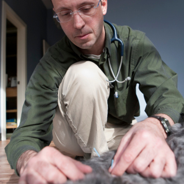
 Healing Paws was founded by Dr. Jeff Corey, DVM, in 2005 as Rhode Island’s first and only comprehensive rehabilitation center with a veterinarian on staff. When Dr. Corey opened Armory Animal Hospital in 2016, Healing Paws was incorporated into the new practice as a complement and continuation to the preventative and surgical veterinary care that he and his staff provide. Today Dr. Corey and his skilled staff heal animal patients from all over southern New England through innovative treatments based on established medical principles – all within a friendly and soothing environment.

Benefits of Veterinary Rehabilitation
Just like physical therapy for humans, veterinary rehabilitation can:
- Reduce recovery time after injury and/or surgery
- Decrease pain and inflammation
- Increase range of motion to help with easier movement during daily activity
- Lead to a longer and better quality of life for your pet
- Aid in weight loss for overweight and elderly patients
- Increase muscle tone and cardiovascular health
These are just some of the most common benefits that rehabilitation can bring to a pet. And for the devoted family, adding more years to their pet’s life – and more life in their years – is a benefit beyond measure.
Veterinary Rehabilitation Services
At Armory Animal Hospital’s Healing Paws Veterinary Rehabilitation Center, we are committed to improving your pet’s health and quality of life using these specialized rehabilitative therapies…
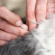 Acupuncture provides pain relief as well as a general sense of wellness, and promotes healing through neuromodulation. The main indications for acupuncture in the rehabilitation setting are arthritis, recovery from neurologic injury, and post-op from a neurological or orthopedic procedure. Conditions and injuries that can benefit from acupuncture treatment include degenerative joint disease (underlying arthritis), underlying neurological problems such as following disc surgery or hindlimb weakness, and, to a lesser extent, GI and skin issues.
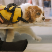 The buoyancy of warm water and the movement of the treadmill combine to reduce the impact on painful joints and help increase the patient’s mobility. Some examples of how hydro-treadmill therapy may be used are after orthopedic or neurologic surgery, to aid in weight loss, or for athletic conditioning.
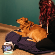 Laser energy is absorbed by the individual cells stimulating them to produce proteins necessary for tissue repair as well as endorphins that help in pain relief. Laser therapy is highly recommended for surgical sites and injury locations. Conditions and injuries that can benefit from therapeutic laser treatment include arthritis, soft tissue injury, trigger points, degenerative myelopathy, and for post-surgical recovery and rehabilitation.
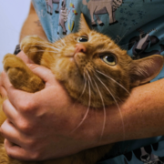 Stretching and corrective movements are used to enhance mobility and promote joint health. Conditions and injuries that can benefit from range of motion exercises include muscle contracture, fibrotic myopathy, neurological problems, generalized pain syndromes, and retraining cats and dogs with decreased mobility.
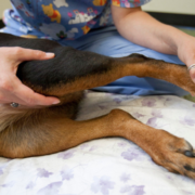 Gentle soft tissue manipulation is used to relax muscles and offer pain relief. Conditions and injuries that can benefit from massage therapy include generalized weakness, muscle atony and for post-surgical or post-athletic eventing recovery.
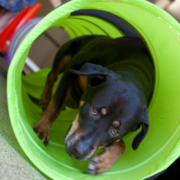 These are exercises performed in the clinic that help strengthen muscles as well as improve the patient’s spatial awareness. Exercises utilizing the physio-ball and wobble board are often employed with a variety of orthopedic and neurologic conditions. Strengthening and proprioceptive exercises are useful in cases where the animal needs to be retrained to walk, for downer dogs, and for any condition where decreased stamina is an issue.
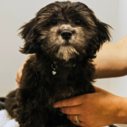 Tailored to each individual pet’s needs, this may include massage, range of motion exercises, and thermo (hot and cold) therapy that the client can do at home. Assuming they are amenable to a hands-on approach to rehab with their owner, dogs with almost every condition can benefit from these individualized programs.

Conditions That Can Be Treated with Veterinary Rehabilitation
All pets recovering from surgery or with conditions causing varying levels of impaired mobility, muscle strength, and pain can benefit from an individualized rehabilitation program. With Healing Paws’ skilled care, pets heal faster and more completely. Here are just some of the conditions that can be improved with rehabilitation:
The Anterior Cruciate Ligament attaches the femur to the tibia (“shinbone”) preventing excessive motion between these two bones and keeping the joint stable. Over-extension of the knee joint may tear this ligament allowing the two bones to slide back and forth causing pain, lameness, and instability.
Excessive movement over a period of time leads to arthritis and pain. Overweight dogs are most susceptible due to the excess pressure created on the joints. Conservative medical therapy initially using anti-inflammatory drugs may allow healing if the ligaments are merely stretched instead of being torn. Complete rest is essential for any chance of healing to occur. If the ligament is actually torn, even partially torn, surgical repair will be needed to form new ligaments and tighten the joint giving stability needed for normal movement and activity.
Degenerative myelopathy is a progressive disease of undetermined cause that affects the spinal cord. It results in a loss of coordination of the hind legs, which progresses to weakness and then to paralysis of the hindquarters. What happens is that the structures within the spinal cord that are responsible for nerve impulses degenerate. In degenerative myelopathy, the myelin (the insulation around the nerve fibers) and the axons (the nerve fibers that carry signals to the muscles) are affected. While these changes can happen anywhere along the spinal cord, they usually happen in the lower back.
Typically, degenerative myelopathy isn’t seen in dogs under the age of five. The degeneration occurs slowly over a period of several months. Often the first signs noticed are difficulty getting up in the hind quarters. This awkwardness is most noticeable when the dog walks on a smooth surface. However, as the disease progresses, the dog becomes uncoordinated and will scuff or drag its rear feet, causing excessive wearing of the toenails.
Sometimes one side is more noticeably uncoordinated than the other. The disease can either wax and wane episodically or progress steadily. It usually takes a few months to a year after onset for a dog to become unable to walk.
There is no known cause for this disorder, although a genetic basis is presumed in German Shepherds. Genetic, nutritional, and immune factors have been suggested, but none have been scientifically proven. While this is mostly a disease of middle-aged to older German Shepherds, German Shepherd mixes, Siberian Huskies, Collies, Corgis, and other breeds can also be affected. It is not thought to cause pain or discomfort because the spinal cord axons have no way to feel pain. Usually, affected dogs can still urinate and defecate on their own until the very late stages.
A DNA test is now available for use by veterinarians, breeders, and pet owners. This test is available through the OFA (Orthopedic Foundation for Animals).
Elbow dysplasia is the most common cause of front limb lameness in the young dog, especially of the larger breeds. Dysplasia comes from the Greek dys, (abnormal) and plassein (to form). Thus, dysplasia refers to abnormal development, in this case, of the elbow joint.
The elbow is formed from the meeting of three bones: the humerus, which is the boney support of the upper limb from the shoulder to the elbow; the ulna, which runs from the elbow to the paw along the back of the limb; and the radius, which supports the major weight-bearing along the front of the lower limb. All three of these bones need to grow and develop normally and at the same rate such that they fit perfectly at the elbow. If there are any abnormalities along these lines or if the cartilage lining the elbow joint does not form properly then “dysplasia” or abnormal formation is the result.
Elbow dysplasia can take several different forms. Specifically, ununited anconeal process (UAP), fragmented medial coronoid process (FMCP), osteochondritis dessicans of the medial humeral condyle (OCD), ununited medial epicondyle (UME), and elbow incongruity all qualify as types of elbow dysplasia that can be present individually or in combination. While all of the variations are distinct and probably develop in different ways, they have in common that they produce loose pieces of bone and/or cartilage within the joint that act as irritants much as a pebble does in your shoe! All of these variations also have in common that they are primary problems that invariably lead to the secondary development of arthritis within the elbow. The term “arthritis” simply describes inflammation within a joint. The longer an elbow joint is ill-fitting or irregular, the more arthritis forms.
While traumatic episodes may affect the development of the elbow joint, the vast majority of elbow dysplasia cases are genetic in origin.
Predisposed breeds include the Bearded Collie, Bernese Mountain Dog, Chow Chow, German Shepherd, Golden Retriever, Labrador Retriever, Newfoundland, Rottweiler, St. Bernard and Bassett Hound.
To understand FCE, one has to understand some anatomy of the vertebral column. The vertebral column consists of numerous small bones called vertebrae that are linked together by special joints called intervertebral discs. The discs are similar to the joints that connect arm or leg bones together in many ways. They allow flexibility between vertebrae so that one can arch or twist one’s back voluntarily just as one can flex and extend a knee or elbow.
But the discs are unique as well. A joint of the appendicular skeleton, say a knee or elbow, has a capsule that secretes a lubricating fluid. The bones are capped with smooth cartilage to facilitate frictionless gliding as the surfaces move during flexion and extension. The disc is nothing like this. It is more like a cushion between the end plates of the vertebrae. It is round (hence the name disc) and fibrous on the outside with a soft gelatinous inside to absorb the forces to which the bones are exposed. This jelly-like material inside is called the nucleus pulposus and it is the is material that makes up the fibrocartilaginous embolus.
The vertebral column provides a bony protective case around the vulnerable spinal cord. The spinal cord is the cable of nerve connections that transmits messages to and from the brain and controls the reflexes of the body. The spinal cord is fed by a network of spinal arteries. In FCE, somehow the material from the nucleus pulposus enters the arterial system and is carried to the spinal cord where it causes a blood vessel obstruction: an embolism. This area of the spinal cord actually dies. The process is not painful but generally recovery is not likely. Whatever neurologic loss has occurred within the first 24 hours is likely to be permanent (though the condition does not get progressively worse).
There are many theories of how disc material might gain access to the arterial blood supply but no one really knows how this happens.
Most FCE dogs are young adults between the ages of 3 and 6 years. In one study, 61% were presented for evaluation after some kind exercise injury or trauma. There may be a yelp at the time of the trauma but the injury is generally not painful. There is about a 50:50 chance that the lumbar area of the spinal cord will be affected, which means only the rear legs will be involved. Because the embolism is not generally a symmetrical event, both left and right may not be equally affected. Along with large breed dogs, some people believe the Miniature Schnauzer is also at increased risk.
The term dysplasia means abnormal growth, thus hip dysplasia means abnormal growth or development of the hips. Hip dysplasia occurs during the growing phase of a puppy, usually a large breed puppy, and essentially refers to a poor fit of the ball and socket nature of the hip.
The normal hip consists of the femoral head (which is round like a ball and connects the femur to the pelvis), the acetabulum (the socket of the pelvis), and the fibrous joint capsule and lubricating fluid that make up the joint. The bones (femoral head and acetabulum) are coated with smooth cartilage so that motion is nearly frictionless and the bones glide smoothly across each other’s surface. When a dog has hip dysplasia, the ball and socket do not fit smoothly. The socket is flattened and the ball is not held tightly in place, thus allowing for some slipping. This makes for an unstable joint and the body’s attempts to stabilize the joint only end up yielding arthritis.
There are two sets of patients typically affected by hip dysplasia. The first group is the adolescent dog, typically 6 to 18 months of age. These dogs may be brought to the vet’s office for signs of discomfort. Radiographs are taken and hip dysplasia is discovered. However, many dogs who would have similar radiographs will not be in pain and thus will not end up coming to the vet for an evaluation. These dogs show up as elderly dogs, after they have been walking on their poorly formed hips for many years. After many years, bony build up along the margins of the socket, mineralization of the joint capsule, cartilage wear, and inflammatory change in the joint (i.e., degenerative arthritis) has become painful and now the dog comes to the vet for an evaluation. Physical therapy and massage are also important and helpful in non-surgical joint therapy.
The bones of the backbone that protect the spinal cord are called vertebrae. Soft cushions are located between these bones, which serve as “shock absorbers” protecting the very delicate nerves that lie within the spinal column. These cushions are called discs. (For more details about the spine, see description for FCE above)
The disc may be damaged from an injury, such as jumping off furniture, resulting in the condition called a ‘slipped disc.’ In this condition, the disc has been forced out of its normal location and pushes against the spinal cord itself causing pressure on the nerves. Pressure results in pain, weakness, lack of coordination, and sometimes paralysis of the legs, bladder, and rectum. Disc protrusion against the spinal cord can also result from a deterioration of the disc as the pet ages or arthritic changes within the bone itself.
Disc disease can occur anywhere along the spinal canal. “Pinched nerves” in the neck area are usually very painful and may cause front leg lameness. The pet often is presented with a reluctance to move the head up and down, usually keeping the head tucked low to the ground. Lesions further down the spinal column cause varying signs depending on the particular nerves compressed by the involved disc. All four legs can be affected in severe cases. Dogs that are overweight are at increased risk.
Obesity has become an extremely important health problem in the Western world, not just for humans but for dogs and cats as well. Obesity in pets is associated with joint problems, diabetes mellitus, respiratory compromise, and decreased life span; recent estimations suggest that up to 35% of dogs and cats in the U.S. suffer from obesity.
A common justification for over-feeding treats is that a pet deserves a higher quality of life as a trade-off for longevity. While this might on some level makes sense (after all, a pet munching on a treat is certainly getting a great deal of satisfaction from doing so), the other consequences do not make for higher life quality in the big picture. Here are some of the problems that obese animals must contend with while they are not enjoying their treats and table scraps: decreased lifespan, increased risk of diabetes, and worsened arthritis due to excessive weight.
The medical term for a dislocation of the kneecap. It is most often seen in the smaller breeds and may be inherited. Many Poodles, Chihuahuas, and other small breeds inherit a very shallow groove on the femur in which the kneecap must ride. Any injury resulting in stretching or twisting of the knee causes the ligaments to tear allowing instability of the patella. It usually is displaced to the inside of the leg and often may “pop” back into the proper location when the leg is straightened, taking pressure off the joint. The leg usually appears to be turned inward when viewed from the rear of the dog. Many times, both rear legs are affected. It is often possible to push the affected patella in and out of the proper position. Surgery is often necessary if lameness persists to prevent later arthritis. Surgery involves deepening the notch in the femur where it should remain positioned and tightening of the various ligaments.
explore more
PREVENTATIVE CARE
Consultations, vaccinations, check-ups, microchipping and more
Contact & Connect
ARMORY ANIMAL HOSPITAL & HEALING PAWS REHABILITATION
1275 Westminster Street
Providence, RI 02909
Phone: 401-648-0009
Fax: 401-453-7245
HOURS
Monday: 8AM – 4:30PM
Tuesday: 8AM – 4:30PM
Wednesday: 8AM – 4:30PM
Thursday: 8AM – 4:30PM
Friday: 8AM-4:30
Saturday and Sunday: Closed
Closed during lunch from 12-1 everyday
